
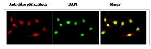
Description cMyc is a proto-oncogene, which is overexpressed in a wide range of human cancers. Myc gene encodes a transcription factor that regulates a great number of genes through binding on Enhancer Box sequences (E-boxes) and recruiting histone acetyltransferase. It can also act as a transcriptional repressor. It regulates cell growth, apoptosis, differentiation and stem cell self-renewal. Previous studies on the phosphorylation of c-Myc have suggested functional association between phosphorylation at Thr58/Ser62 by glycogen synthase kinase 3, cyclin dependent kinase, ERK2 and C-Jun N terminal Kinase (JNK), cell proliferation and cell cycle regulation. Phosphorylation at Ser62 is required for Ras-induced stabilization and is prerequisite for phosphorylation at Thr58 for its degradation (ref.1). Applications Western blot (~1 μg/ml, Fig.1) Immunofluorescence staining (0.5~1 μg/ml, Fig.2) Immunohistochemistry (5 μg/ml, Perform heat mediated antigen retrieval with citrate buffer pH 6 before formalin treated paraffin embedded sectioning) Flow cytometry (Use 1 μg for 106 cells) Indirect ELISA (Assay dependent concentration) Specification Antigen: Synthetic peptide containing phospho-Ser62 of cMyc Isotype: Mouse IgG2b (κ) Form: Purified monoclonal antibody (IgG) 1mg/ml in PBS, 50% glycerol Reactivity: Human. Expected to react with mouse and rat from the sequence identity. Storage: Ship 4°C and store at -20°C (long period, -70℃) Data Link UniProtKB/Swiss-Prot P01106 (MYC_HUMAN) Fig. 1 Identification of cMyc protein whose Ser62 is phosphorylated by Western blot. Samples: Crude cell extracts of AGS (gastric adenocarcinoma) cells. Scr: scrambled siRNA was introduced into the cells as a negative control. Neg. Control: Negative control siRNA from Qiagen was transfected. Myc1: siRNA for cMyc was transfected. The data was provided by Drs. A. Khanna and J. Westermark of University of Tampere. Fig. 2. Immunofluorescence staining of cMyc phosho-Ser62 in nuclei of HeLa cells. HeLa cells were fixed with 4% paraformaldehyde overnight, permealized with 0.25% Triton X-100 in PBS for 10 min. Cells were blocked prior to staining with 1/2,000 diluted anti-cMyc p62 antibody in 1% BSA in PBS at 4℃ overnight. Cells were stained with a secondary antibody, goat anti-mouse IgG conjugated with Alex 488, at 1/1,000 dilution in 1% BSA for 1 hr at room temperature. Nucleus (DNA) was stained with DAPI. References Sears R et al. Genes Dev 14:2501-2514 (2000) PMID: 11018017 Junttila MR et al. Cell 130: 51-62 (2007) PMID: 17632056 Khanna A et al. J Natl Cancer Inst 101: 793-805 (2009) PMID: 19470954
Related products
JACOBY CUES
$ 99.86
$ 96.08
CUETEC CUES
$ 99.86